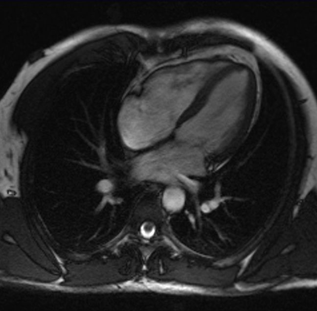
Cardiac magnetic resonance imaging (MRI) uses a strong magnetic field, radio waves and a high-end computer to create very detailed still and moving pictures of the structures of the heart (cardiac chambers, valves, heart muscle, vessels) and surrounding region. Numerous images are taken from different angles of the heart and heart vessels throughout the scan. With the help of the recorded images, it is possible to see and measure how the heart is functioning and how the blood is flowing through the valves and vessels. This advanced diagnostic technology can provide detailed information on the type and severity of several heart diseases.
At Medicare Hospital we use SIEMENS MAGNETOM Aera MRI scanner for fast and reliable scans and the outmost image quality and resolution.


