Skin lesion removal

What is a skin lesion and how does it develop?

Abnormal growths and parts of the skin which appear different to their environment are called skin lesions. They can be classified as either primary or secondary. Primary skin lesions such as birthmarks, acne and moles can either be present at birth or acquired over a person’s lifetime.

Secondary skin lesions, however, develop as a result of irritation to primary skin lesions. For example, if someone scratches a mole until it bleeds, the resulting lesion is a secondary one. Chronic conditions (poor circulation, diabetes), infections, allergic reactions or sensivities can also lead to the development of skin lesion.

What types of skin lesions exist?

There are many different types of skin lesions. The most frequest ones are the following:

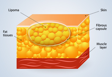
Lipoma

A lipoma is a slow-growing, fatty lump which develops in the soft tissue between the skin and the underlying muscle layer. A lipoma feels like a soft rubbery bulge and usually isn’t tender, moves readily with slight finger pressure.

Lipomas are usually detected in middle age and are most often located on the torso, arms, or thighs. Some people have more than one lipoma. Lipomas are classified as tumors but they are not cancerous and usually do not require any treatment. However if your lipoma bothers you, is painful or is growing, you may want to have it removed.

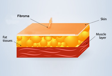
Fibroma

Fibromas are benign tumors made up of fibrous or connective tissue cells. They can grow in any part of the body. When they grow on the skin they have a brownish colour and are soft to the touch. They can often be found in the armpits, neck and groin area. It is not known exactly what causes fibromas to appear, but it is suspected that both hormonal and genetic factors may play a role in their development. There are several types of fibroma including soft (contains few cells), hard (contains many cells), keloid (develops from healed wounds), cystic (lymph vessels among the connective tissue) and angiofibroma (blood vessels among the connective tissue).

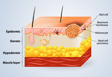
Basalioma

Basalioma (also known as basal cell carcinoma or BCC) is the most common type of skin cancer. While basalioma has a very low metastatic risk and hardly ever results in death, this tumor can cause significant disfigurement by invading the surrounding tissue.

It often appears as a waxy bump, though it can take other forms. Basal cell carcinoma occurs most often on areas of the skin which are often exposed to the sun, such as the face and neck.

Skin pigment disorders

Skin color is determined by melanin, a pigment produced by specialized cells in the skin (melanocytes). A person’s skin color. depends on the amount and type of melanin secreted.

Pigmentary disorder is an umbrella term which includes hyperpigmentary disorders (darkening of the skin) and hypopigmentary disorders (lighter than normal skin color), the most common disorders being melasma (hyperpigmentary) and vitiligo (hypopigmentary).

Sebaceous cysts

Sebaceous cysts are benign tumors beneath the surface of the skin. They originate from sebaceous glands and contain sebum – primarily composed of triglycerides, wax esters, squalene, and free fatty acids – secreted by those glands. These cysts are formed when the opening of a sebaceous gland gets clogged for some reason and sebum is unable to find a way out causing the gland to swell. Most sebaceous cysts are found right beneath the skin, usually on the face or scalp. Their size vary from about 1cm to several centimeters.

When does a skin lesion need to be removed?

Many skin lesions do not require removal at all while some are removed for cosmetic reasons. Another reason for the excision of skin lesions is to fully remove skin cancers or other abnormalities which have the potential to become cancerous in the future if left untreated. In these cases surgery is used to prevent the tumor from metastasizing (spreading) to the surrounding tissue and other parts of the body. Skin lesions are sometimes removed for diagnostic purposes, or to prevent irritation, infection and inflammation.

How is a skin lesion removed?

A variety of techniques is used to remove skin lesions. The particular technique selected will depend on a number of factors such as the type of the lesion, its location, and the patient’s ability to tolerate the procedure. The aim of these surgeries is to completely remove the lesion and clean the surrounding area. The operation usually takes 15 to 45 minutes, duration largely depends on the type and extent of the lesion.

What are the risks and benefits of the operation?

In most cases abnormal skin growths can be removed quickly and effectively without any significant risk of bleeding or infection. Usually a specimen of the removed tissue is sent to a laboratory and placed under closer analysis called histopathology to determine what type of cells it is made up of, and whether there are any cancer cells present.

Some scarring is to be expected as it is not possible to cut the skin without leaving some sort of mark. Your dermatologist will excise the lesion and repair the wound in a way that leaves the smallest possible scar. Unfortunately some people’s wounds do not heal as they should, they may develop something called keloid or hypertrophic scarring.

Am I a good candidate for skin lesion removal?

In order to diagnose a skin lesion, your dermatologist will want to conduct a full physical exam. Treatment is based on the underlying cause(s) of your skin lesions. The doctor will take into account the type of lesion, personal medical history, and previous treatments.

How to prepare for the surgery?

Before the surgery please list all medication you are currently taking, including vitamins and supplements. You will be given a detailed medical questionnaire and requested to inform your medical team about any allergies, as well as current and past illnesses.

What to expect after a skin lesion has been removed?

Keep the wound dry and follow your doctor’s instructions regarding aftercare. It is very important to to be gentle with that part of your body and to keep it clean at all times. A degree of pinkness and tenderness to touch around the wound edges is normal but if the wound becomes increasingly red or painful, consult your dermatologist promptly – it could be a sign of infection in which case you may need a course of antibiotics.

Why is regular follow-up important after surgery?

Initially the scar will be red and slightly raised but it will gradually shrink. Within a few months the color of the skin will become lighter and appear quite similar to the surrounding tissue. It is recommended to have a follow-up appoitment 7 to 14 days after surgery to make sure that the wound is healing as it should.


Dear Customers,

we would like to inform you that our Clinics will be closed on August 20th and 21st.

Medicover
close-link


Dear Customers,

we would like to inform you that our Clinics will be closed on August 20th and 21st.

Medicover
close-link