What is the difference between cervical conization and cryotherapy?

What is the difference between cervical conization and cryotherapy?

15-06-2018

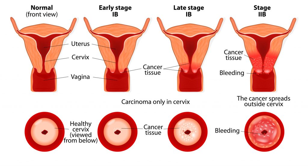
Cervical cancer is the second most common cancer among women after breast cancer. Most often it develops around the age of 40. Despite modern prevention, today 1 in 25 000 women die from cervical cancer in the WHO European Region. Taking into consideration these data we cannot emphasize enough that regular gynecological exams can easily prevent the formation of cervical cancer.

The surface of the cervix is easily accessible during the gynecological examination, from which smear can be taken for further analysis. Since cervical cancer develops over the years, there is a cancer-preventing condition that helps to prevent the disease before the malignant lesion develops.

There are many methods for preventing cervical cancer, all of them are used by doctors at a different stage of the lesion. In our current blog post we compare conization and cryotherapy.

Tissue excision procedures – conization

There are basically two methods of conic and loop excision, but they also have different versions. The Greek-Latin name of cone-shaped excision of the cervix is conization.

Usually, a lancet is used for the surgery, also known as cold-knife conization. We distinguish between heat-consuming incisions methods (electric-knife), but surgery can also be performed by laser (laser conization).

The surgery is done under general anesthesia. During the procedure you lay in the position well-known from gynecological examinations; the surgeon reaches your cervix through your vagina so there are no unnecessary cuts on your abdomen. During conisation, we remove the entire transitional part, but if it is too large, only the affected part is cut off by the surgeon, the remaining part of the transitional area may be treated by a tissue destruction process (e.g., cryotherapy).

Tissue destruction methods – cryotherapy

The tissue destruction procedures are performed in outpatient care by local anesthesia preceded by a colposcopically controlled tissue sample (biopsy) and its histological examination.

Various methods are used, including freezing, heat treatment, and cold coagulation.

Cryotherapy (freezing) is using high degree cold to destroy tissue lesions. The operating temperature of the freezer units is approximate -80 °C, which freezes the cells quickly.

Freezing is usually used twice for three minutes with a five-minute break. Choosing a suitable head for the size of the lesion is extremely important: it is essential that the tip of the head covers the whole surface of the lesion, or even overlaps with the 3-5 mm.

The success of cryotherapy depends primarily on the extent and not the severity of the lesion; large ones can be more difficult to handle. The method is painless and very simple, with no major complications. The only consequence of the intervention is the colorful, water-like flow that lasts for 3-4 weeks, resulting from the destruction of the dead tissue.

As the treatment takes place in outpatient care, the patient can leave the hospital shortly after the cryotherapy procedure and may be in the work the next day.


Dear Customers,

we would like to inform you that our Clinics will be closed on August 20th and 21st.

Medicover
close-link


Dear Customers,

we would like to inform you that our Clinics will be closed on August 20th and 21st.

Medicover
close-link